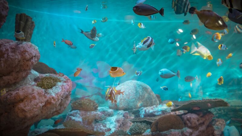
Aquarium Parasite Identification Guide: Visual Diagnosis
Spotting trouble early can mean the difference between a quick treatment and a devastating tank wipe. This aquarium parasite identification guide from Gensou Aquascaping at 5 Everton Park, Singapore, will help you visually diagnose the most common parasites that affect freshwater and marine fish. Recognising what you are dealing with is the first step toward choosing the right treatment and saving your livestock.
White Spots: Ich and Velvet
Ichthyophthirius multifiliis, commonly called ich or white spot disease, is the parasite most hobbyists encounter first. The telltale sign is grain-of-salt-sized white cysts scattered across fins, body, and gills. Each cyst is roughly 0.5-1 mm in diameter. Fish often flash against hardscape and breathe rapidly as the parasite irritates gill tissue.
Velvet disease, caused by Piscinoodinium pillulare in freshwater or Amyloodinium ocellatum in marine tanks, looks subtly different. Instead of distinct white dots, you will notice a fine, dusty gold or rust-coloured coating — most visible when you shine a torch at an angle across the fish’s body. Velvet tends to kill faster than ich because it attacks the gills aggressively. In Singapore’s warm ambient temperatures of 28-32°C, both parasites cycle rapidly, so treatment must begin the moment symptoms appear.
Anchor Worm and Fish Lice
Anchor worm (Lernaea) is a crustacean parasite that embeds its head into the fish’s tissue, leaving a visible thread-like body dangling outside. You may also see a red, inflamed wound at the attachment point. These parasites are often introduced with wild-caught fish or live food purchased from local shops around Serangoon North or Thomson.
Argulus, or fish lice, are flat, disc-shaped parasites around 5-10 mm wide that cling to the skin. They are easier to spot than most parasites — look for a translucent, rounded shape that moves when disturbed. Both anchor worms and fish lice can be removed manually with tweezers under sedation, but the tank still needs treatment to eliminate larvae.
Gill and Skin Flukes
Flukes are flatworms belonging to the genera Dactylogyrus (gill flukes) and Gyrodactylus (skin flukes). They are microscopic, so you cannot see them directly. Instead, watch for behavioural clues: rapid gill movement, flashing, clamped fins, and a thin mucus coating on the body. A skin scrape examined under a basic microscope at 40x magnification will confirm their presence — the worms appear as tiny, wriggling hooks.
Gill flukes are egg-layers, meaning treatment must be repeated to catch hatching larvae. Skin flukes give live birth, so a single round of praziquantel is often sufficient. Knowing which type you are dealing with directly affects your dosing schedule.
Internal Parasites: Worms and Flagellates
White, stringy faeces are the classic sign of internal parasites. Hexamita and Spironucleus flagellates cause hole-in-the-head disease in cichlids, producing pitting erosion around the lateral line and head. Camallanus worms are unmistakable — thin, red-brown threads protrude from the fish’s vent, sometimes waving visibly.
Tapeworms are harder to spot externally. A bloated abdomen combined with weight loss despite normal feeding is suspicious. If you see flat, segmented white sections in the faeces, tapeworm is likely. Treatment with levamisole or fenbendazole, available from local aquarium pharmacies or via Shopee, targets these effectively.
Identifying Parasites Versus Bacterial or Fungal Infections
Not every white growth is a parasite. Cotton-wool-like tufts are usually fungal (Saprolegnia), while red streaks on fins and body point to bacterial haemorrhagic septicaemia. A useful rule of thumb: parasites tend to produce symmetrical, repeating patterns — dots, dust, or visible organisms — while bacterial infections create irregular patches, ulcers, or fin erosion. Fungal growths are fluffy and localised, often appearing on wounds or dead eggs.
When in doubt, treat the most dangerous possibility first. In Singapore’s warm water, bacterial secondary infections develop quickly on parasite-damaged tissue, so combining antiparasitic treatment with a mild antibacterial like methylene blue is common practice.
Tools for Visual Diagnosis at Home
A basic USB microscope (available for $30-$60 on Lazada) is invaluable. Scrape a small amount of mucus from the fish’s body using a blunt coverslip, place it on a slide with a drop of tank water, and examine at 40-100x. You will be able to identify flukes, Trichodina (spinning disc-shaped ciliates), and Epistylis (stalked colonial parasites often mistaken for ich).
Photograph any external symptoms with your phone’s macro mode before starting treatment. This gives you a visual record for comparison and is useful if you need to consult experienced hobbyists on local forums or seek advice from a specialist at Gensou Aquascaping.
Quarantine and Prevention Protocol
Every new fish should spend a minimum of two weeks in a quarantine tank. A simple 40-litre container with a sponge filter, heater (rarely needed in Singapore unless treating cold-water species), and PVC pipe for hiding is enough. Prophylactic treatment with praziquantel for flukes and a general antiparasitic covers the most common hitchhikers.
Maintaining stable water parameters — 0 ppm ammonia, 0 ppm nitrite, and nitrate below 20 ppm — reduces stress and keeps your fish’s immune system strong. Healthy fish resist parasites far better than stressed ones. An aquarium parasite identification guide is most useful before you need it, so familiarise yourself with these visual cues now rather than scrambling during an outbreak.